





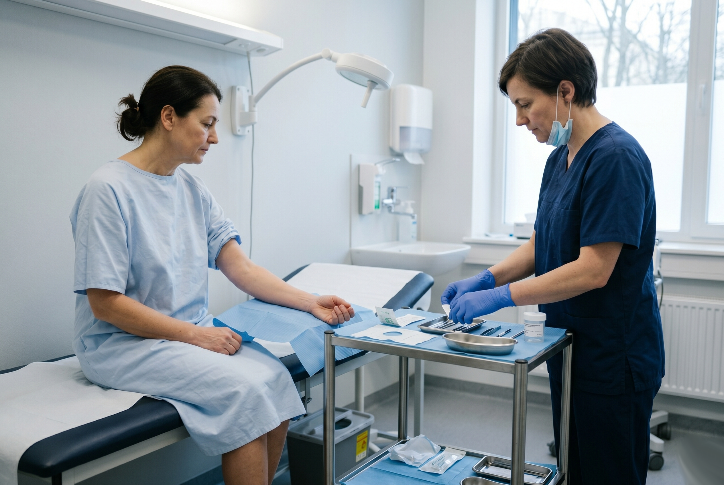
There is no blood test that confirms eosinophilic fasciitis. There is no imaging finding specific enough to close the case on its own. What settles the diagnosis in most patients is a piece of tissue — and specifically, a piece of tissue taken deep enough to include the layer the disease is named after[1].
This article describes what that procedure involves, why the depth matters so much, and how to read a report that comes back ambiguous.
Why a biopsy at all
The diagnostic criteria in common use pair one major criterion — symmetric, plate-like induration of the limbs sparing the fingers and toes — with a set of minor criteria, of which the histopathological picture is one[1]. In practice, most centres want the tissue.
The reason is differential diagnosis rather than confirmation for its own sake. Systemic sclerosis, morphea, eosinophilia-myalgia syndrome, nephrogenic systemic fibrosis and chronic graft-versus-host disease can all produce hardened skin over a limb. They begin in different layers, and the only way to see which layer is involved is to sample all of them at once.
Some centres do diagnose without a biopsy when the clinical picture is classic and the minor criteria are clearly met — particularly when contrast MRI shows the typical fascial pattern. Whether tissue is required is a decision that depends on the centre’s experience and on how much diagnostic doubt remains.
What “full-thickness” actually means
The procedure is a full-thickness skin-to-muscle biopsy. It is surgical, not a needle sample, and it takes a wedge of tissue that passes through every layer in sequence:
- Epidermis and dermis
- Subcutaneous fat, with its vessels and septa
- Superficial fascia
- Deep fascia — the layer the diagnosis turns on
- The superficial layer of the underlying muscle
Only the last two layers answer the question being asked. Everything above them is sampled because the pathologist needs to see where the abnormality starts and stops, not because those layers are themselves diagnostic.
The same limb, two different procedures. A punch reaches the dermis; the diagnosis lives several centimetres deeper. This difference accounts for a large share of first biopsies reported as negative.
The comparison explains one of the most common diagnostic dead ends in eosinophilic fasciitis. A punch biopsy is quick, done under local anaesthetic in an outpatient room, and leaves almost no scar — but it samples the dermis, and in classic M35.4 the dermis can look close to normal while the deep fascia several centimetres below is thickened many times over. A report of 'no significant abnormality' from a punch is therefore not evidence against the diagnosis; it is evidence that the wrong depth was sampled. The full-thickness procedure costs more, needs an operating setting and leaves a real scar, but it is the only version that reaches the layer the disease is named after.
Pinal-Fernandez 2014 · Onajin 2022
Before the procedure
Two things happen before the scalpel that have more influence on the result than the surgery itself.
Site selection. Contrast MRI is the standard way of choosing where to cut. It shows fascial thickening, T2 hyperintensity and post-gadolinium enhancement, and it marks the area where those changes are most pronounced[4]. Biopsying a clinically affected but radiologically quiet area is one of the routes to a false-negative result.
Steroid timing. Eosinophils disappear from tissue quickly once glucocorticoids are started, even while the disease itself continues. A biopsy taken after treatment has begun can show fascial thickening and inflammation without the eosinophils that give the disease its name.
If a biopsy is being planned and steroid treatment has already started, that sequence is worth raising explicitly with the treating team, because it changes how the report should be interpreted rather than whether the procedure is worth doing.
What the pathologist looks for
The report is not a yes/no verdict. It is a description of four features, and their combination is what carries the weight:
- Fascial thickening — commonly described as two to ten times normal
- Inflammatory infiltrate within the fascia — lymphocytes, plasma cells and macrophages, with eosinophils present in roughly half to sixty per cent of samples and more often in early disease
- Thickened collagen bundles — with active fibroblast activity early on, and mature, hyalinised fibrosis in longer-standing cases
- Extension into neighbouring layers — the infiltrate reaching the subcutaneous tissue above and the perimysium of the muscle below
Notably, the muscle itself is usually normal. That is one of the features that separates M35.4 from the inflammatory myopathies, where the muscle signal and muscle histology are both abnormal.
When the report says “no eosinophils”
This is the sentence that causes the most confusion, and it does not carry the meaning it appears to.
The absence of eosinophils does not exclude the diagnosis. They vanish from tissue after steroid exposure, and they are frequently sparse or absent in the chronic fibrotic phase even in untreated patients[2]. The diagnosis rests on the whole picture — clinical presentation plus the fascial changes — not on whether one cell type happened to be present in one sample on one day.
The historical irony is that the disease was named for a laboratory finding present in the first described cases[3] that turns out to be absent in a substantial minority of patients.
The biopsy in context
Against the other investigations, the full-thickness biopsy is the most sensitive single test — but the chart below is best read as an argument for combining tests rather than for relying on any one of them.
Proportion of patients in whom each investigation shows typical changes. Biopsy leads, but the supporting tests are what make the clinical picture add up.
Full-thickness fascial biopsy leads the list at roughly 95 % because it samples the layer where the disease actually sits. Contrast MRI follows at 80–90 % and is the best non-invasive option — strong enough to guide the biopsy site and to track treatment response, not specific enough to replace histology. Peripheral eosinophilia is present in around 90 % of pooled published patients but is entirely non-specific on its own, and it is suppressed within days of starting steroids. Hypergammaglobulinaemia sits around two thirds. Musculoskeletal ultrasound trails at about a quarter — useful for follow-up and for excluding alternatives such as deep-vein thrombosis, inadequate as a diagnostic step. No single row here is sufficient; the diagnosis is assembled from several of them at once.
Pinal-Fernandez 2014 · 2025 systematic review (n = 597)
Recovery and the wait
The wound is closed with sutures and the scar is real — several centimetres, usually on the forearm, thigh or calf. Ordinary wound care applies, and the limb is generally used normally within days.
The longer part is the wait for the report. Histopathology on a full-thickness sample involves fixation, sectioning and staining, and turnaround measured in weeks rather than days is common. It is worth knowing in advance that this gap exists, because for many patients it lands exactly in the period when symptoms are still progressing.
TIP: We recommend that you consult your doctor. Thank you!
References
- Pinal-Fernandez I, Selva-O'Callaghan A, Grau JM. Diagnosis and classification of eosinophilic fasciitis. Autoimmun Rev · 13(4-5):379-382. 2014. PubMed · 24424187
- Lebeaux D, Sène D. Eosinophilic fasciitis (Shulman disease). Best Pract Res Clin Rheumatol · 26(4):449-458. 2012. PubMed · 23040360
- Shulman LE. Diffuse fasciitis with eosinophilia: a new syndrome?. Trans Assoc Am Physicians · 88:70-86. 1975. PubMed · 1224441
- Moulton SJ, Kransdorf MJ, Ginsburg WW, Abril A, Persellin S. Eosinophilic fasciitis: spectrum of MRI findings. AJR Am J Roentgenol · 184(3):975-978. 2005. DOI · 10.2214/ajr.184.3.01840975
- Naschitz JE. Clinical guide to eosinophilic fasciitis: straddling dermatology and rheumatology. Expert Rev Clin Immunol · 18(7):649-651. 2022. PubMed · 35575016 DOI · 10.1080/1744666X.2022.2078309